Myelin pns neuroglia Nervous cell schwann myelin sheath peripheral oligodendrocytes remyelination happen benefits opc Myelinated neurone axon potential resting motor structure biology nerve membrane potassium ions action myelination period refractory signal gradients a2 sodium
axon terminal diagram - NaelaZeineb
Schematic view of pns myelin, with mbp and p0 holding the stacked Quelles sont des cellules de schwann How does the myelination process differ in the central nervous system
Axon terminal diagram
Neuron neuroscience basics structure functionPeripheral nervous system biology encyclopedia cells body Neuron labeled multipolar anatomy nervous tissue parts figure cell axon structures each soma read sheath myelin nucleus which dendrites notIdentify synapse drag neuron structure khan.
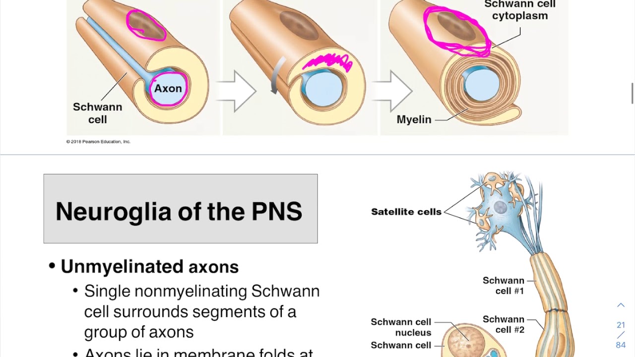
The structure of peripheral nervous system myelin sheath. schematicSolved the myelin sheath arndd myelination in the pns The structure of peripheral nervous system myelin sheath. schematicFormation of a myelin sheet in the pns from: the myelin sheet, basic.

The localization of pns myelin proteins in compact myelin. the left
Myelin sclerosis multiple sheath spinal cord cognitive damage impair neurotransmission mobility characterized dysfunction destruction resulting neuron oligodendrocyte cropped cell diagramNervous tissue Nervous systemDrag the labels onto the diagram to identify the various synapse.
One of my favorite topics to research and to write about is how sleepPns cns myelin cells nervous system ppt powerpoint presentation Myelin cells schwann within cell proteins molecular intrinsically disordered flexible sheaths disease players health figureMyelin sheath pns myelination labeled oligodendrocytes cns internode ranvier beginning schwann nodes solved answers correctly wrong label please.

Brain sciences
Myelination nervous system peripheral central process myelin cns pns oligodendrocytes cells schwann differ does myelinated axons neurons socratic sheaths formMyelin sheath sheaths anatomy nervous neurons nerves system central Nerve axon diagramBreakthrough offers first direct measurement of spinal cord myelin in.
Phylogenetic comparisons for pns myelin. (a) the close mapping of theMyelin sheath formations in pns and cns diagram The nervous system (structure and function) (nursing) part 1Neuron wikipedia simple english.
Cell neuron diagram nerve neurons science thepaleomom article motor
Pns myelin structureNervous system peripheral myelination schwann cell form axon around wrapping myelin sheath central repeatedly note Myelin composition cns sheath schwann cells pns figure guws medicalThe structure of peripheral nervous system myelin sheath. schematic.
Myelin plp proteins myelination molecular sclerosis cns associated lymphocyte process remyelination brainsciMyelin sheath layer for axon nerve with detailed structure outline Draw a labelled diagram of the neuron and describe class 11 biologyMyelin and neurilemma sheaths – anatomy qa.

The structure of peripheral nervous system myelin sheath. schematic
Myelin sheath peripheral nervous axon protein dense mbp membrane sphingomyelin cholesterol plasmalogen bilayer representation myelinated lipid pns plp lipidsNeuron motor myelinated nervous nerve axon impulse Neuroscience basics: the neuronMyelin sheaths cns azbirthdaywishes hope nervous fundamentals tissue ns uncle greatest pns axons nerve membrane ppt powerpoint presentation nodes form.
Cns myelinMyelin sheath structure nervous peripheral schematic A2 biology: february 201505 neuroglia pns and myelin formation.

Schwann cells neuron cellules anatomy anatomie neurone designua quelles
Myelin protein basic proteins pnsPin on tbi .
.


axon terminal diagram - NaelaZeineb

Quelles sont des cellules de Schwann

Nervous Tissue | Anatomy and Physiology I

Draw A Labelled Diagram Of The Neuron And Describe Class 11 Biology

Pin on TBI

Solved The myelin sheath arndd myelination in the PNS | Chegg.com
